PPT The Lower Limb PowerPoint Presentation, free download ID2968685
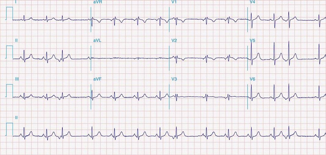
The S1S2S3 pattern, in conjunction with right-dominant forces on a 12-lead electrocardiogram including a tall R-wave in lead V 1 (R:S >1), deep S waves in the left precordial leads V 5 and V 6 (R:S <1), QRS interval <120 ms, and right atrial enlargement (P-wave in lead II >2.5 mm), is highly specific for right ventricular dysfunction with pulmon.
PPT Cardiovascular Step 1 Review PowerPoint Presentation, free download ID2253521
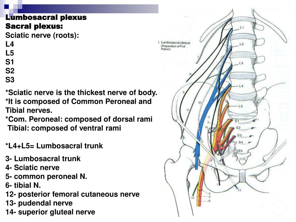
The sacral plexus begins as the anterior fibres of the spinal nerves S1, S2, S3, and S4. They are joined by the 4th and 5th lumbar roots, which combine to form the lumbosacral trunk. This descends into the pelvis to meet the sacral roots as they emerge from the spinal cord. Fig 1 - The spinal cord outflow at each vertebral level.
Heart Sounds Concise Medical Knowledge
What Are The Four Heart Sounds? Medical Editor: William C. Shiel Jr., MD, FACP, FACR Definition Function Lub Dub Sounds What are heart sounds? Using a stethoscope to assess different sounds the heart makes is an important diagnostic tool.

Pin on heart sounds
The sacral plexus is a network of nerves formed by the lumbosacral trunk (L4, L5) and sacral spinal nerves (S1 - S4). The sacral plexus is located on the posterior pelvic wall, posterior to the internal iliac vessels and ureter, and anterior to the piriformis muscle.

Microscopic aspect of S1, S2 and S3 foliations moving from S to N a)... Download Scientific
This may be because the three heart sounds — S1, S2, and S3 — in quick succession create a cadence, or rhythm, like a galloping horse. Additionally, the heart sounds follow a similar galloping.

Heart Sounds S1 S2 S3 S4 Pearson+ Channels
A combination of 5 nerve roots that exit from inside the lower lumbar and upper sacral spine—L4, L5, S1, S2, and S3—forms the sciatic nerve. These 5 nerves group together deep in the buttock, near the front surface of the piriformis muscle, and combine to form the single large, thick sciatic nerve. 1 Davis D, Vasudevan A. Sciatica.
The arrows point out the S1S2S3 foramina for transsacral block. Download Scientific Diagram
S1 refers to the first sacral bone, S2 to the second sacral bone, and so on. S1 is at the top and S5 is towards the bottom. Each number corresponds with the nerves in that part of the spinal cord. S1 nerves affect the hips and groin. S2 nerves affect the back of the thighs. S3 nerves affect the medial buttock area.

Auscultation area listening positions for S1, S2, S3, and S4 heart... Download Scientific Diagram
Superior gluteal nerve, formed by sections of L4, L5, and S1; Inferior gluteal nerve, formed by sections of L5, S1, and S2; Sciatic nerve, which is the largest nerve of the sacral plexus and among the largest nerves in the body, formed by sections of L4, L5, S1, S2, and S3; The common fibular nerve (formed by L4 through S2) and tibial nerves (formed by L4 through S3) are branches of the.

Heart sounds s1 s2 s3 s4 Auscultation areas lub dub sound of the heart why these areas
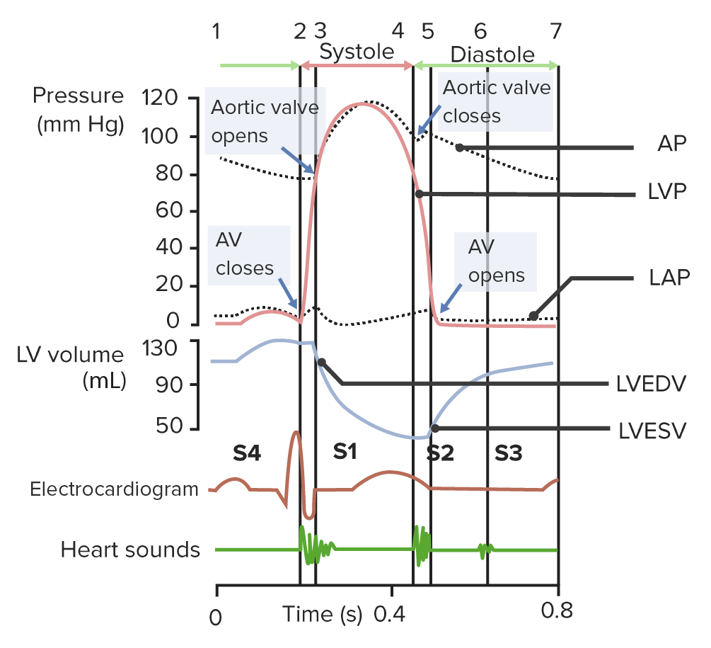
Bruits and Hums of the Head and Neck. Heart sounds are created from blood flowing through the heart chambers as the cardiac valves open and close during the cardiac cycle. Vibrations of these structures from the blood flow create audible sounds — the more turbulent the blood flow, the more vibrations that get created.

Motor duty and it's types explained.S1,S2,S3,S4 meaning. YouTube
The sacral spinal nerve 3 (S3) is a spinal nerve of the sacral segment.. It originates from the spinal column from below the 3rd body of the sacrum.. Sacrum, showing bodies in center. Muscles. S3 supplies many muscles, either directly or through nerves originating from S3. They are not innervated with S3 as single origin, but partly by S3 and partly by other spinal nerves.

Medical knowledge, Tricuspid valve, Mitral valve
Essentially the difference between a S1,S2 and S3 safety shoe are the degrees of protection the boots provide to the wearer. Please check out this guide to learn more about which safety boot is right for you. safety shoes manufacturer One professional safety shoes company that produce different type of safety shoes and work boots in China.
Atlas of Noninvasive Imaging Basicmedical Key
Burch and de Pasquale defined S1S2S3 as S waves in the leads I-III with S2 > S3 [15]. In our study, only 252 individuals had S2 ≥ S3 and either S1S2S3-I or S1S2S3-II. The S1S2S3 pattern is not a typical finding of left anterior fascicular block, where, by definition, the S wave in lead III is deeper than the S wave in lead II,.

PPT Cardiovascular System PowerPoint Presentation, free download ID2925304
What the heck? Instead, these bikes use Specialized's new S-Sizing system: S1, S2, S3, S4, S5, and S6. So how does S-Sizing work? It might seem confusing at first, but it's actually quite clever.. At 5'8, I could comfortably ride an S2, S3, or S4. But since I want to go as fast as possible on chunky, high-speed local trails, I would.
Anhängen an Enttäuschung so wie das heart tones Fest Wissenschaft Kapok
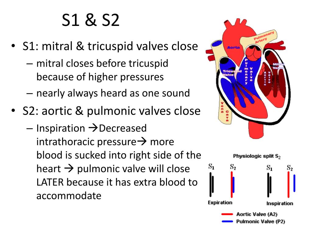
by Ali Al-Hadithi | 26 Jan, 2021 Cardiology Cardiovascular Examination S1 and S2 Heart Sounds, Extra Heart Sounds Login Basics of Heart Sounds - S1 and S2 heart sounds There are 2 main heart sounds that can be heard during auscultation: S 1 and S 2, also affectionately known as 'lub' and 'dub' respectively.

Nursing Assessment of the Cardiovascular System
Act as a shock absorber for the spine and control the transmission of forces from the lower body into the spine, such as gravitational forces and forces transmitted upward during standing or walking. 2 Wong M, Sinkler MA, Kiel J. Anatomy, Abdomen and Pelvis, Sacroiliac Joint. [Updated 2020 Aug 10]. In: StatPearls [Internet].

Heart sounds Best Nursing Schools, Nursing School Studying
The S1, S2, S3 Syndrome in Chronic Pulmonary Disease E. A. was a 50 -year-old white man with severe far-advanced emphysema. His electrocardiogram is an excellent example of the S 1, S 2, S 3 syndrome. Note that there is a prominent S wave in Leads 1, 2, and 3 and the S waves are equal in duration and magnitude to the preceding R waves.